Germination

...and she says: "What a life!"

August 10, 2011

Day 8

THE GRAND ROUND

I never knew what a grand round was until today. It's basically like a session where all physicians are present and they discuss about a medical case. And today, they discussed about this condition called Dermatomyositis. It is a disease that involves the skin and muscles of the body.

Patient was 21 years old when she was diagnosed with hypertension and was given beta-blocker to bring down her hypertension. After 5 years she stopped the drug as she felt well. However, she started the feel lethargic and rashes started to appear on her skin. She couldn't do simple exercises and had to quit her job. She was then referred to this hospital from Sibu and initially they thought that she was having heart failure. So when they sent her to Sarawak International Medical Centre, she was diagnosed with no cardiac events! However, her CPK enzyme was high (CPK enzymes are released from muscles so it is not specific to the heart). So then she was sent to SGH for rheumatology unit and they started to identify signs and symptoms that she presented. One of the most prominent one was Gottron's papules. There is also proximal muscle weakness. And so the story went on and she was diagnosed with dermatomyositis. She was then screened for any malignancy. Apparently, in a few studies, it was indicated that there is a higher chance of patient who has dematomyositis to have nasopharyngeal carcinoma. And, an important point to note, dermatomyositis is closely related to malignancy.

Gottron's papules- macular

A differential diagnosis for dermatomyositis is polymyositis. However, the differences between this two diseases are:

1. Dermatomyositis is caused by antigen-antibody complex that accumulates at the tissues that causes the manifestations of the disease.

Polymyositis is a delayed hypersensitivity reaction.

2. Dermatomyositis is a strong malignant condition.

Polymyositis has a low connection with malignancy.


Remember that when doing a confirmative diagnostic test, muscle biopsy should always be taken from the most normal muscle not those which has atrophied AND muscle biopsy should NOT be done after electromyography(EMG) as during EMG, the muscles are excited and is traumatic to the muscle which gives an inaccurate test result.

This is an autoimmune disease and is common in women. So, to know more about the disease, one should read up on it. This post is just a personal note and reference so do not fully rely on what is written here.

Labels:

August 9, 2011

Motivational quote

"When you have a bad day because you’re tired, stressed, overworked, and underappreciated, never forget that things are much worse for the person on the cold end of stethoscope. Your day may be lousy, but you don’t have pancreatic cancer." - member of BMJ's editorial board

Labels:

Day 7

OH MY GOSH! Being at home is SO BORING. I can't wait for 2pm to come so I can go to the hospital, at least learn something. I hope 2 hours in the paed's cardiac clinic will be fruitful today. Boredom is hard to bear. I should have brought a book back from KL to read... >.<

August 8, 2011

Day 6

Coronary artery bypass graft (CABG) - Open heart surgery baby! :D


I was lucky to be able to enter the OT to see an open heart surgery for CABG.

This patient had Percutaneous Coronary Intervention (stenting) done before but then it's not working anymore and that artery that received PCI had narrowed down. So the surgeons have to replace the narrowed artery and create a collateral branch for it for collateral perfusion.

Patient came in at 8am and got prep for surgery.

First, neck line was set up. They had trouble setting it on the left side so they changed it to the right. While at it, the surgeon marked the surface marking for harvesting the great saphenous vein at the right inner thigh.

Second, urinary catherization.

Third, skin preparation. Basically, sterilization. I guess it's to avoid any pathogens to enter the body through the incisions they were gonna make later.

And of coz, syringes with medications, surgery equipments which I couldn't name and monitoring equipments to monitor the patient's time to time status.

At last, they started the surgery at 9.40am.

There were 4 surgeons doing the surgery. 2 harvesting the great saphenous vein, 1 (main surgeon) harvesting the internal mammary artery and 2 (main surgeon and another surgeon) doing the CABG.

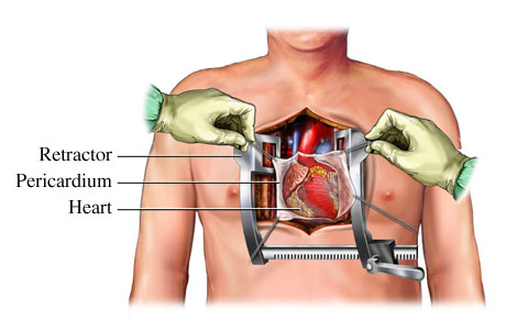
The main surgeon made an incision on the midline of the chest- sternotomy. This will then expose the sternum. The surgeon then used an equipment which worked like a saw to cut open the sternum into half to reach the thoracic cavity. The saw the left lung from the place where i was seeing.

Great saphenous vein harvesting:
The surgeons made 2 separate incisions on the right inner thigh. One is above the knee, and the other one is about 3 fingers above the first incision. When they incised the thigh and initially exposed the great saphenous vein, it was a bit BLUISH! :D and then it turned slightly red. The great saphenous vein is quite big. It's like a white colour wire when they took it out. Diameter is around 3mm? The length they harvested is about 10cm i think. I am not very sure. They ligated the smaller veins that previously drained into it and removed it from the inner thigh.

Internal mammary artery harvesting:

MAN! This is so cool! This is like the happiest day of my life. WHY? I got to see the heart and lungs IN SITU! with the sternum cut open to expose them.The chest was opened up using a retractor. The was chest wide open and u can actually see all the layers. and there, when the surgeon was harvesting the left internal mammary vein, the innermost layer of the chest just above the lungs. I COULD SEE THE LEFT LUNG EXPAND. It's such a historical moment-for me. I could clearly see that the left lung has 2 lobes. However, I didn't get to see the lingula. URGH!

After the harvesting, the surgeons who were responsible for taking out the great saphenous vein put the vein in the kidney dish and sew up the incisions. While the main surgeon and another surgeon worked their way through the pericardium to reach the heart.

They opened the pericardial sac and push it aside, exposing the right atrium (I could see the right auricle too), the right ventricle, the aorta and the pulmonary artery. :D The heart is enveloped by a layer of thick fat tissue. The surgeons then lifted the heart and also started the bypass grafting. Amazing! The surgeon used this super fine thread to stitch the super fine vein when the end of it was a little bit torn. SALUTE! They have this special glasses so I guess that helped them to magnify the fine broken part of the vein when they were stitching. After that the remaining main process was to anastomose the internal mammary artery and the great saphenous vein on the surface of the heart.

At the end of the procedure, which was almost 4 hours in the OT, CABG was finally done and the surgeon used this thick sternal wire to stitch the sternum back together and close the incision.

Open heart surgery is always an impressive surgery to me.

What I learnt:

1. In the OT, never turn ur back against the sterile equipment.

2. Stand one foot away from the sterile equipment.

3. The low temperature, around 16 degree celcius is compulsory to reduce the risk of infectious pathogens being active and infect the opened chest.

4. CABG- they use great saphenous vein and internal mammary artery.


Labels:

August 5, 2011

[cont. II] Day 5

CVS history taking report

Personal details

Chief complaint

History of presenting illness

Background /Past medical history/ Surgical history

Drug history: ALLERGIES, medications

Social history


Clinical examination


BP, HR

Systemic review (compulsory)

CVS, RS, GIT, CNS

Examination for the fingers: the splinter hemorrhages can be accentuated by shining a torch on the finger examined.

Auscultation:

1. Basic auscultaion: Mitral , Tricuspid, Aortic and Pulmonary

2. Accentuation of murmurs: Ask patient to breath IN to check for RIGHT side murmur and breath OUT for LEFT side murmur.

3. Ask patient to lean forward for aortic regurgitation murmur and lean to the left side for mitral stenosis murmur.


* cardiac risk factors are to be included in PMH:
- modifiable: dyslipidemia, diabetes mellitus, smoking, alcohol
- non-modifiable: 1st degree relative who have a history of heart disease

Labels:

[cont. I] Day 5

There are so many things that I learnt today.

What I learnt:

1. Angina: the description by patient can be not exactly what was describe in the book. For example, patients may describe it as SOB.

So, assume the patient is correct at what he is describing, check for

- heart failure symptoms, COPD to rule out SOB
- then try to identify if it is of heart origin
- check with echocardiogram, ecg...


2. Diabetic patients treatment progress:

- Metformin: increase sensitivity of tissues to insulin

- Sulphonylurea: stimulate pancreatic function to increase insulin secretion (used in patient with failing pancreas)

- Insulin injection: when pancreas cannot produce enough insulin


3. Dialysis patient:

- Renal failure causes the body to retain fluid as it is not excreting. This can cause the blood pressure to increase due to fluid retention. During dialysis, the fluid is removed from the body and with this, the blood pressure will drop. If not monitored properly, patient's blood pressure will drop and cause the patient to faint.

- Antihypertensive drug is withheld before dialysis procedure.

4. Pregnant patient who are at a young age and have chest pain and SOB during labour, we should consider the following conditions that might happen but not CAD (age is incompatible with this cardiac event):

- Suspect Rheumatic Heart Disease (RHD), mitral valve abnormality (MR or MS)

- Suspect pulmonary embolism due to DVT: pregnant uterus can compress the inferior vena cava and impair the return of the blood causing stasis in the vessels of lower limbs.

Test that can be carried out:

~ D-dimer test: breakdown of clot. In pregnant woman, it can be slightly elevated esp in those who underwent C-section. This test can be slightly non-specific.

~ Doppler studies: on the vessels of lower limbs to indicate signs of thrombosis.

5. Read up on thyrotoxicosis and Graves disease.


Labels:

Day 5

Today is a very productive day as I learnt a lot.

I went to the specialist clinic today and sat in in the consultation room. Dr. Ong attended 8 patients. I learnt a lot from him. Most of the patients are follow up or referred patients. The interesting ones are the one I have listed here.

A male patient came in with a history of coronary arterial disease (CAD) and a MI. Due to the scarring from MI, he needed to undergo ablation therapy or he will get ventricular tachycardia. He is also planned for implanted cardiac defibrillator (ICD) too.

A female patient had chest pain and SOB during labour. She was given Caeserean section to deliver her baby. Due to her condition during the labour, a cardiac event must be rule out in case anything happens. So she was referred to the Cardiology department. During history-taking, the patient is too young to be suspected of CAD, so the other causes for chest pain should be considered. What are differential diagnoses? Refer here (point 4).

After the clinic session, I went to the Cardiothoracic ward (CTW) and Dr Khiew was there. He wanted me to present the case which I have clerked 2 days ago. When I wanted to present, he stopped me and told me how case presentation is done. It's like a revision and in depth CVS history taking. After that, he brought me for bedside teaching. I was told to do physical examination on a real patient. I feel like digging a hole and HIDE MYSELF. URGH! so many mistakes and the worst parts were: I didn't introduce myself and state my purpose of interview; I didn't ask if the patient was in PAIN! DAMN...HE WAS A REAL PATIENT, YVONNE! >.<


Labels:

August 4, 2011

Day 4

I got to enter the cardiac lab to see how angiogram is done.

Prep the patient by cleaning the sites of incision with iodine+alcohol.

Inject local anaesthetic- Lignocaine.

Insert catheter into the radial artery.

Guide wire is used to guide the tube into the aortic root.

The contrast dye is injected to examine if there is any abnormality in the coronary arteries.

A patient had 3 arteries narrowed. Another had severely calcified coronary arteries.

Example of an angiogram:


What I learnt:

Left coronary artery is divided into Left Anterior Descending and Left Circumflex.

Right coronary artery gives the Posterior Descending Artery

Labels:

August 3, 2011

Day 3

The patient who had MI and was put on peritoneal dialysis passed away. RIP.

I went for ward round with the whole team of consultant, cardiologist, MO and HO.

The cardiologist gathered 4 of us- the students, in front of CCU and made us go get patient's history plus physical examination. We have to present it to him tomorrow morning. So we all went to CTW to hunt for patients. THIS IS WHAT MEDICAL STUDENTS DO. urgh!

I paired up with Nora. We went to talk to this female patient-CSN.

Basically, CSN was admitted with swelling of lower limbs and shortness of breath. She experienced orthopnea and PND so she could only sit up while sleeping for the past 3 months. She went to the doctor's 3 months ago and was given frusemide to be taken twice daily each time a tablet. When admitted on 1/7/2011 she was diagnosed with pulmonary edema, decompensated heart failure due to the failing biosynthetic valves she got 10 years ago. Upon auscultation she had pansystolic murmur.

What I learnt today:

I went to INR clinic today and saw that many patients were on warfarin. A doctor taught us these:

Cardiology:

1. Pseudo- mitral stenosis: A pansystolic murmur can indicate mitral regurgitation or aortic stenosis. However, when there is aortic regurgitation,which is a diastolic murmur, there will be a leak from the aortic valves, this will cause a pressure to push the leaflet of the mitral valve towards the left atrium. This will then cause the mitral valve to partially closed causing pansystolic murmur. So, there will be a mix diastolic+pansytolic murmur.

2. Cardiology has 4 parts: a. Acute coronary syndrome (STEMI,NSTEMI,USA) b. Heart failure c. Arrhythmias d. Congenital Heart Disease

3. Drugs for STEMI:

A- aspirin, ACEi, ARB
B- beta-blocker
C- calcium-channel blocker
D- diuretics
S- streptokinase, statins

Labels:

August 2, 2011

Day 2

The Head of Department of Cardiology was on leave so I was passed to another cardiologist. I went for ward rounds with the Consultant(Dr Yew), Cardiologist(Dr Khiew), MOs and HOs plus one new friend who is from Royal College of Surgeons Ireland (RCSI).

After ward round, I went back to CCU and basically I stayed there for the rest of the day.

Most memorable patients:

Patient 1: Indian male with MI with subsequent episodes of AF and VT.

Patient 2: Native male with 3rd degree heart block. He was sent for pacing wire procedure later in the morning. I got to read his ECG and was able to pick out the abnormality- there weres P waves seen but there was no QRS complex.

Patient3: Chinese male with heart failure due to graft blocked. Patient had 2 open heart surgery for 2 CABGs. One of the CABGs was blocked, causing his heart to fail, renal shutdown as a consequence and pulmonary edema. He was put on peritoneal dialysis. I witnessed how femoral vein catheter was inserted.

What I learnt:

When doing incision for any procedure involving the area of the groin, remember not to do it above the inguinal ligament. As the inferior epigastric artery is nearby, if punctured will cause hematoma which will bleed into the peritoneal cavity unnoticed.

Please know the antibiotics well!

What I revised:

Anatomy relations of femoral nerve, femoral artery, femoral vein and femoral canal (lateral to medial).

External iliac artery gives off the inferior epigastric artery.


Labels:

August 1, 2011

Day 1- SGH

7.05am I arrived at Sarawak General Hospital.

Walked down the hall towards the administration office. The guard said it opened at 8am.

8.00am Entered the administration office and registered. I was told that I needed a passport size photo for the temporary ID. I filled in the form and chose 3 departments to attach to for the next 3 weeks - Cardiology, General Surgery and General Medicine.

General Surgery FTW!

I know...Dr. Derek Shepherd is a neurosurgeon.


2.oopm I handed in my passport photo and got my ID done at 4pm.

On a side note, ppl can be SO LAZY! It's just pasting the photo on the card, get it signed by the person in charge and lamination. WHY MUST I WAIT TILL THE NEXT DAY? AND 8AM IS TOO EARLY FOR YOU TO PASS IT TO ME IF I COME BACK THE NEXT DAY. -.-"

I demanded my ID to be done by today so I can report for duty on time tmr orelse I am gonna miss a day again.

THESE PPL!ISH!


Labels: